價格:免費
更新日期:2018-12-10
檔案大小:7.6 MB
目前版本:3.0
版本需求:需要 iOS 12.1 或以上版本。與 iPhone、iPad 及 iPod touch 相容。
支援語言:英語

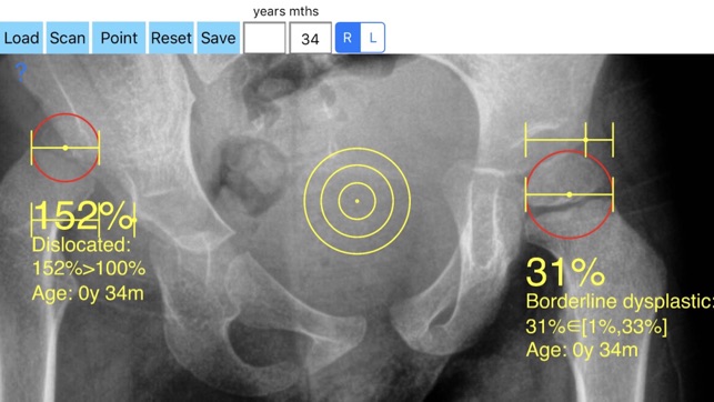
The migration percentage (MP) as described by Reimers is the gold standard method for assessing radiographs especially in children with hip dysplasia which quatitively shows how much femoral head has been dislocated from the ossified acetabular roof.
The Reimer index is calculated as the ratio (alb) x100% where a is the distance measured the lateral border of the femoral head to the Ombredanne-Perkins line and b is the width of the femoral head parallel to Hilgenreiner's line. After the age of 5 years old when the cartilages in hip - especially the triradiate cartilage- are fused, the Reimer index in everyday clinical practise is usually described as the Femoral Extrusion Index. The values are expressed in percentage % of the femoral head that is not covered by the acetabulum and the normal values vary according to the age of the patient. The advantage of this index is its independence of pelvic or thigh rotation and provides a good quantitative estimate of the degree of uncoverage of the femoral head. This cannot be estimated by a decrease in the CE angle of Wiberg.

In a busy everyday practice measuring angles in X-rays in clinical settings it is time consuming and cumbersome. Accessory instruments like protractors, goniometers, well sharped pencils, rulers or even transparent papers must be available. Also after measurement you have to compare the data that you measure with the normal reference values according to patient age, and decide what could be considered normal or pathologic.
The Reimer Index app is medical software aimed for orthopaedic surgeons, providing tools that allow doctors to: -Securely import medical images directly from the camera or stored photos.

-Offers a very convenient way to determine the most accurate possibly way of measuring the percentage of femoral head which is not covered. By the aid of a circular transparent template which help to mark accurately the points of interest and the dynamic graphics who has been especially developed ,by clicking only two points the percentage is calculated. By inputting the age of the patient in the App, the measured values in percentage are compared with values from normal reference database accordingly. In cases the percentage of uncovered femoral head of the hip is beyond the normal range for the relevant age, the hips are categorised as borderline dysplastic or dysplastic ready to subluxate or subluxated ,dysplastic luxated or dislocated .
-Save the planned images, for later review or consultation. All information received from the software output must be clinically reviewed regarding its plausibility before patient treatment! Reimer Index app is indicated for assisting healthcare professionals. Clinical judgment and experience are required to properly use the software. The software is not for primary image interpretation.

The app is a handy tool for an orthopaedic surgeon, radiologist, medical student or resident who wants objectively monitor and determine the severity of dysplasia of the hip. The build-in comparison feature with the normal reference values according to patient age may help decide what could be considered normal or borderline dysplastic or dysplastic and help also judge which hip is subluxate or at risk and help monitor objectively the treatment in time. The app is not a simple goniometer, is an enhanced product which offers the ability to compare all the input data with medical reference database. This feature it is particular useful especially in clinical settings where you need a quick results without losing time in looking for reference data in textbooks.
1.Reimers J. The stability of the hip in children. A radiological study of the results of muscle surgery in cerebral palsy. Acta Orthop Scand Suppl 1980; 184: 1-100

2.Li PL, Ganz R.Morphologic features of congenital acetabular dysplasia: one in six is retroverted. Clin Orthop Relat Res. 2003 Nov;(416):245-53
支援平台:iPhone, iPad
